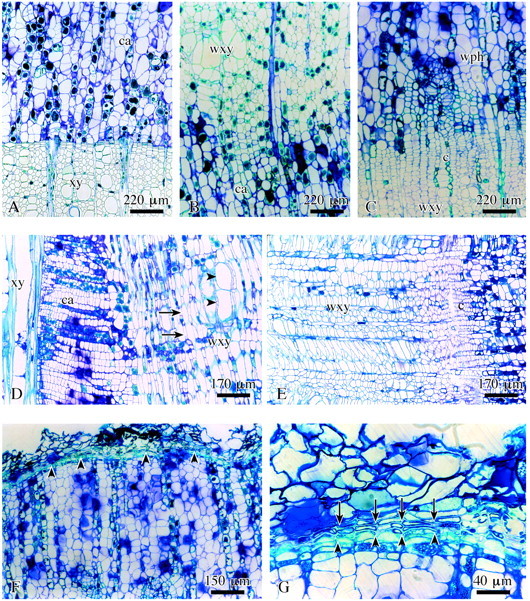
Fig. 6. Structure of completed surface callus in lime. A, Callus cells (ca) close to previous year’s xylem (xy). B, Wound xylem (wxy) adjacent to the callus tissue. C, Wound tissue: xylem, cambium (c), phloem (wph). D, Wound xylem with short fibres (arrows) and vessels (arrowheads) in radial view. E, Wound cambium (c) cells are axially shortened as compared with regular cambium cells. F, Wound periderm formation (arrowheads) completed on the outside of the surface callus. G, Detail of F showing inner phellogen layer (arrowheads) and distinctly suberized phellem cells (arrows) of the wound periderm. A–C, LM; F and G, transverse sections; D and E, radial sections.
