
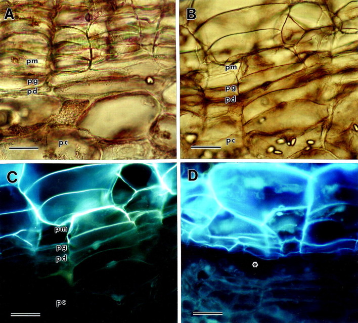
Fig. 1. Light micrographs of potato periderm sections viewed under brightfield or UV epifluorescence (pm, phellem; pg, phellogen; pd, phelloderm; pc, parenchyma underlying the periderm). A, Mature native periderm viewed under brightfield. Bar = 30 µm. B, Immature wound periderm viewed under brightfield. Bar = 60 µm. C, The same immature wound periderm viewed under UV epifluorescence. Note that the autofluorescence of the phellem tissue ends where the phellogen cell layer begins. Bar = 60 µm. D, Excoriated immature wound periderm viewed under UV epifluorescence, demonstrating that wall fracture occurs in the phellogen layer, just under the phellem. The area of the fracture is indicated by an asterisk. Bar = 60 µm.