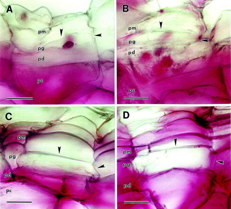
Fig. 4. Ruthenium red staining of wound periderm sections (pm, phellem; pg, phellogen; pd, phelloderm; pc, parenchyma underlying the periderm). Arrowheads indicate phellogen upper tangential and radial cell walls. A, Immature wound periderm. No periderm cells walls are stained, though the walls of the parenchyma cells underneath the periderm are stained intensely. B, Immature wound periderm de‐esterified with 0·1 m Na2CO3 before staining. No periderm cells are stained, although the walls of the parenchyma cells underneath the periderm are stained intensely. C, Mature wound periderm. Phelloderm cell walls are stained, but phellogen cell walls are not stained. Phellem cell wall staining is weak and sporadic. D, Mature wound periderm de‐esterified with 0·1 m Na2CO3 before staining. All three periderm cell types are stained, though staining of phellem and phellogen cell walls is uneven and weak. Bar = 40 µm.
