
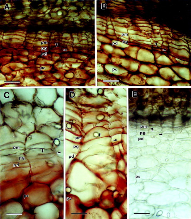
Fig. 5. Peroxidase staining utilizing guaiacol as the substrate. Hydrogen peroxide was added just prior to the guaiacol unless otherwise indicated (pm, phellem; pg, phellogen; pd, phelloderm; pc, parenchyma underlying the periderm). Arrowheads indicate phellogen upper tangential and radial cell walls. A, Immature native periderm. The entire periderm is stained red‐brown, including phellogen cell walls. B, Mature native periderm. The entire periderm is stained red‐brown including phellogen cell walls. C, Immature wound periderm. There is no staining in the phellem or phellogen layers, though the walls of the phelloderm are stained weakly. In contrast, red‐brown staining is intense in the cell walls of the parenchyma underneath the periderm. D, Mature wound periderm. Phellem cell walls are stained red‐brown, but the radial and lower tangential phellogen cell walls are unstained. There is some weak scattered staining in the phelloderm. E, Mature native periderm without hydrogen peroxide added prior to staining. There is no staining, demonstrating the specificity of the reaction for peroxidase (the brown/black colouration in the upper phellem and netting above the phellem is natural colouration and does not represent peroxidase staining). Bar = 60 µm.