
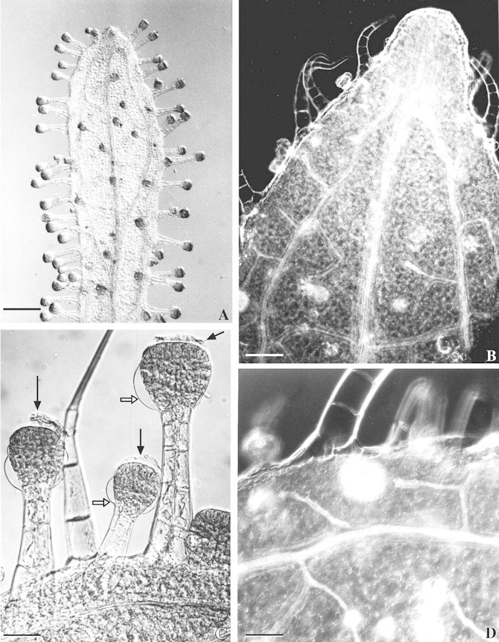
Fig. 2. Vascularization of the outer bracts and long‐stalked glandular hairs of Sigesbeckia jorullensis. A, Outer involucral bract with a large median vein, two lateral veins, smaller veins, and many long‐stalked glands. Bar = 1 mm. B, The three veins fuse to an epithem hydathode. Bar = 200 µm. C, Hairs treated with glycerol‐ethanol and chloral hydrate show the remnants of the cuticle forming the subcuticular space (black arrows) and the girdle between the upper and lower layers of cells, where the cuticle of the lower parts of the head (white arrows) is fixed to the cell wall. Bar = 100 µm. D, Small veins, mainly composed of tracheids, lead to the base of gland cells. Bar = 100 µm.