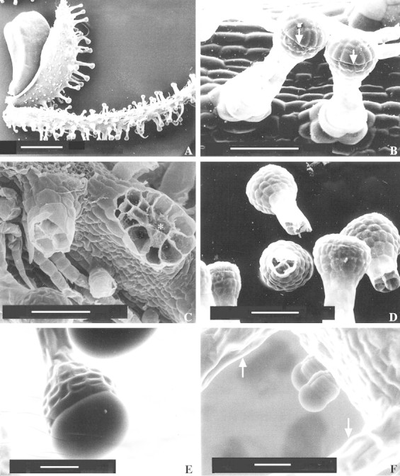
Fig. 3. SEM micrographs of Sigesbeckia jorullensis. A, Fruit dispersion unit consisting of an achene that had been torn out of the receptacle together with the outer and inner involucral bracts. Bar = 1 mm, magnification 22×. B, Young long‐stalked glandular hairs showing the line (arrows) where the cuticle is detached from the wall. Bar = 100 µm, magnification 359×. C, Broken hairs. The base of the hair on the right‐hand side (asterisk) shows 16 cells, whereas the stalk of the hair on the left‐hand side is composed of four cells. Bar = 100 µm, magnification 307×. D, Broken hairs showing four to six stalk cells. Bar = 100 µm, magnification 249×. E and F, ESEM‐mode. E, Long‐stalked hairs without preparation, showing the filled subcuticular space. Bar = 50 µm, magnification 397×. F, Two small ten‐celled hairs and stalks of long hairs (arrows). Bar = 50 µm, magnification 376×.
