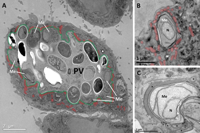
Figure 1.

Host cell mitochondria associate with the parasitophorous vacuole of E. cuniculi.A. Overview of a RK-13 cell infected with a large E. cuniculi PV. The PV (false-coloured in green) encloses a range of different microsporidial stages including meronts and spores (Me, meronts; remaining cells inside PV are spores, recognizable by their discernible cell wall and the ovoid cell shape). Meronts are exclusively located close to the PVM whereas spores differentiate within the body of the PV lumen. Numerous mitochondria (false-coloured in orange) are clustered at the PVM surface preferentially next to meronts.B. Profile of an early meront stage with mitochondria (false-coloured in orange) that ‘cover’ the PVM over large proportions of meront surface.C. Higher magnification of a PV region with a meront bulging into the host cytoplasm. The meront is extensively covered with a mitochondrion that is attached over the full length of its profile. Notice the consistent and close intermembrane spacing between outer mitochondrial membrane and PVM.Me, meront; asterisk, mitochondrion; N, nucleus of meront.
