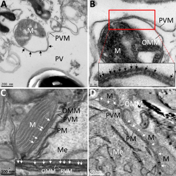
Figure 4.

Binding of host cell mitochondria to the PVM in a broken cell system reveals electron dense bridges.A. Region of an E. cuniculi PV in a broken RK-13 cell. The broken cell appears devoid of granular cytosol and parasite cells as well as host organelles are identifiable. A mitochondrial profile can be seen bound to the PVM (black arrows). This interaction remains intact even when discernible separation of the outer mitochondrial membrane inner membrane has occurred.B. Mitochondrial profile bound to the PVM. Several electron dense bridges (better seen in the higher magnification view) are visible, stretching between the outer mitochondrial membrane and the PVM.C. Mitochondrial binding to the PVM in an intact cell. Close observation of the intermembrane space between the PVM and the OMM reveals electron dense linkages (see white arrows in micrograph and enlargement).D. Snapshot of an electron tomogram showing several mitochondria bound to the PVM surrounding a meront cell. Electron dense bridges can be seen spanning the OMM and PVM (see white arrows).M, mitochondrion; Me, meront; OMM, outer mitochondrial membrane; PV, parasitophorous vacuole; PVM, parasitophorous vacuole membrane.
