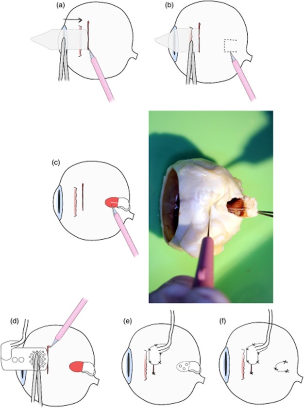
Figure 3.

Procedure to address the issue of posterior ciliary neurovascular bundle (PCNB) obstruction during array insertion. (a) First, a 9-mm full-thickness scleral incision was made, (b) then the suprachoroidal space was probed with a lens glide to find any potential obstruction. (c) If an obstruction was present, a ‘trap door’ scleral incision of 2 × 2 mm was created, and the PCNB was severed.(d) The array was then inserted into the suprachoroidal space. (e) The scleral wound and patch were then sutured. (f) Finally, the ‘trap door wound’ was sutured.
