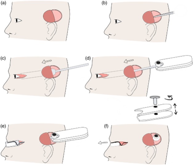
Figure 4.

Pedestal placement and tunnelling. (a) First, an incision site superior to the pinna was made and a full thickness ‘C’ shape flap created on the scalp. (b) Incision was made in temporalis facia, and a tunnel was created between muscle and facia. (c) The tunnelling trocar was passed anterior from ‘C’ shape flap to the lateral canthus to create a tunnel. The temporalis facia was incised lateral to the orbit margin to allow the trocar to pass. (d) The array was placed in the trocar and tunnelled from pinna to the lateral canthus. (e) The lid of trocar was removed. (f) The handle screw was removed, and the handle was separated. The array was then removed from trocar, and the trocar was removed from the head through the lateral canthus wound.
