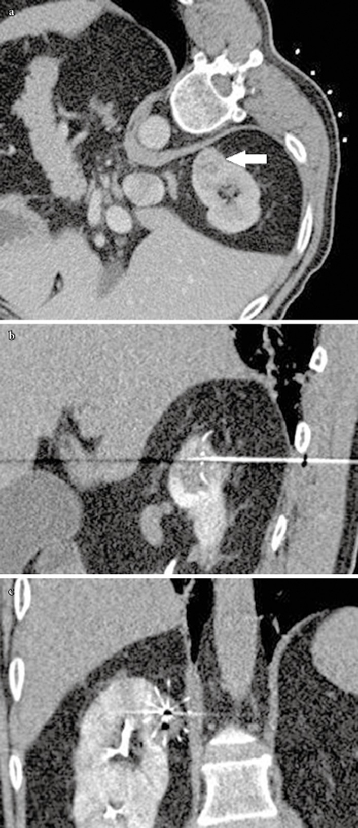
Figure 1.

(A) Pre RFA axial contrast enhanced CT showed a 2.5 cm enhancing renal tumour at the upper pole of the right kidney (white arrow) (B) Sagittal reformatting showed the forward RF burn and (C) Coronal reformatting showed the overall coverage of the tumour by the multi-tines RF electrode.
