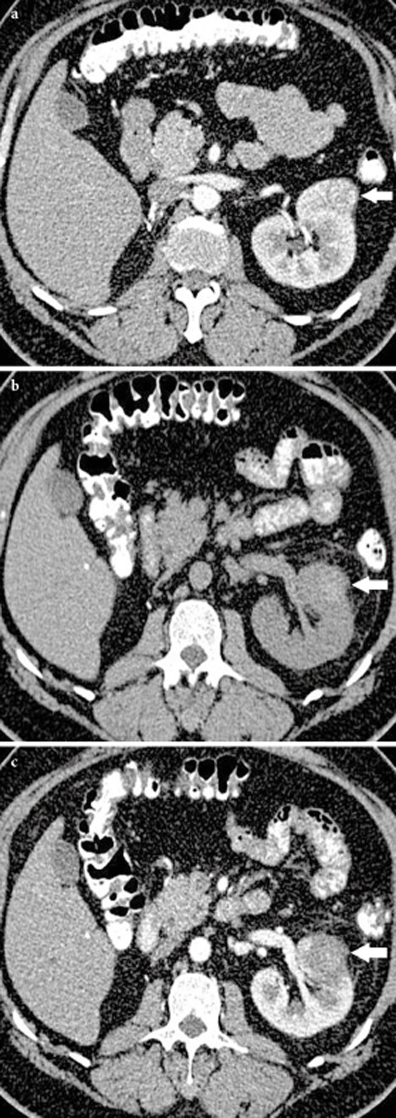
Figure 3.

Axial contrast enhanced CT showed a small enhancing left renal tumour (white arrow) at the anterior cortex of the kidney pre-RFA (A) and the zone of ablation had high attenuation HU post RFA consistent with coagulation necrosis (white arrow) on the unenhanced CT (B) and displayed no enhancement (white arrow) post contrast administration (C).
