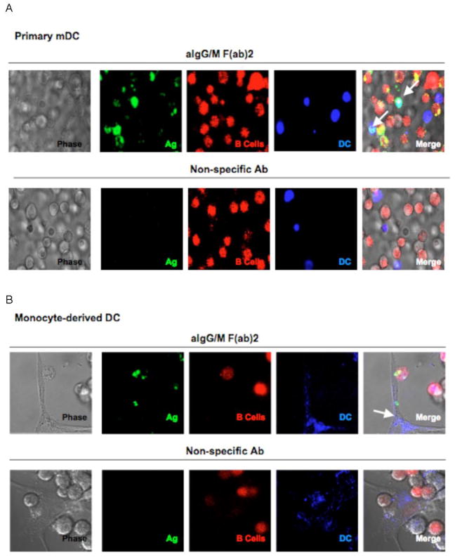
Fig. 2.
Live cell images depict antigen transfer from B cells to dendritic cells. BJAB cells were labeled with CTO and pulsed with antigen. Primary myeloid DCs (a) or monocyte derived DCs (b) were labeled with CTFR and then co-cultured with either aIg (upper panels) or non-specific Ab (lower panels) pulsed B cells in glass bottom dishes. After 18 hr, live cell images were taken by confocal microscopy (600X, 2x zoom; arrows point to DCs that bear antigen).

