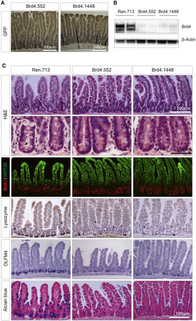
Figure 4. Brd4 Suppression Results in Loss of Intestinal Cellular Diversity.
(A) Immunohistochemical staining showing GFP expression (brown) in intestines of shBrd4 mice on the dox diet. Tissues were counterstained with hematoxylin.
(B) Western blot of Brd4 protein levels in intestinal villi from CAGrtTA3-expressing TtG-Ren.713, TtG-Brd4.552, and TtG-Brd4.1448 mice, maintained on a dox diet for 2 weeks.
(C) Histological and immunohistochemical stains of small intestine sections from CAGrtTA3-expressing TtG-Ren.713, TtG-Brd4.552, and TtG-Brd4.1448 mice. Included are H&E stains (where arrows indicate the location of eosinophilic granules of Paneth cells), immunofluorescent stains for BrdU incorporation (marking proliferating cells) and Keratin 20 (KRT20-differentiated cells), immunohistochemical staining for Lysozyme (Paneth cells), in situ hybridization for Olfm4 (intestinal stem cells), and Alcian blue stains (goblet cells). Scale bars are indicated.

