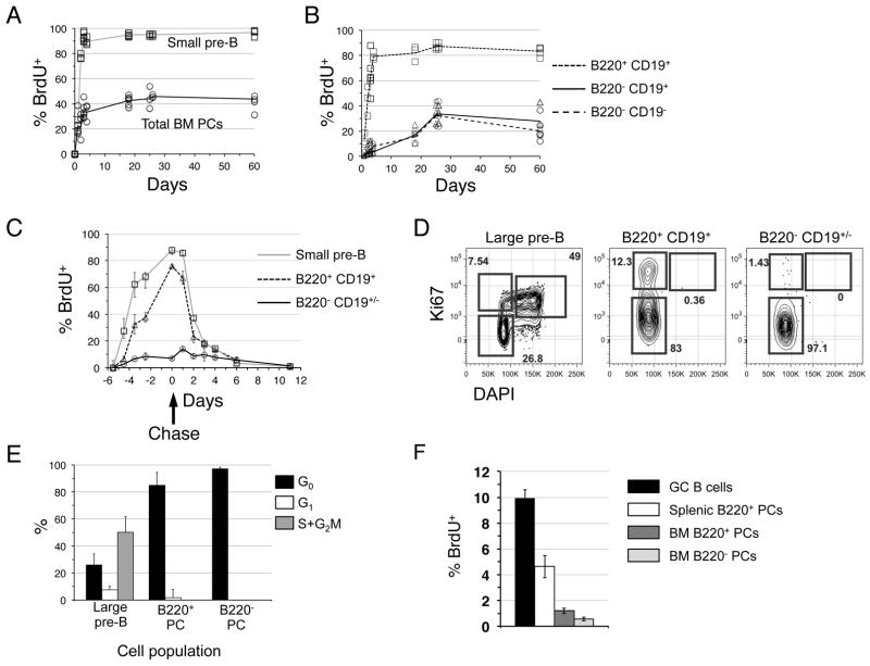
Figure 2. Many BM plasma cells are recently formed.
(A) B6 mice were fed BrdU for the indicated days before determination of the % BrdU+ cells among all Dump− IgD− CD138high BM cells. Small pre-B cells were gated as FSClow B220low AA4+ IgM− cells. Best-trend lines were drawn across the mean % BrdU+ cells for each population using 3–4 mice per time point. (B) The flow cytometric data in (A) were gated as shown in Figure 1B to determine the fraction of BrdU+ cells in the indicated populations at each time point. (C) B6 adults were given BrdU for up to six days, and then given BrdU-free drinking water (chase) for another 12 days. The fraction BrdU+ cells in each BM population was determined as in (A) and (B). (D) BM cells from 12-week old B6 mice were stained to resolve large pre-B cells (FSChigh B220low AA4+ IgM−) or PC subsets based on differential B220 expression, fixed and permeabilized, and then stained with anti-Ki67 antibodies and DAPI. (E) Data summarized for 3 mice using gates shown in (D). Representative of two separate experiments. (F) 8-week old B6 mice were inoculated with BrdU. Sixty minutes later BrdU+ cells were identified among the indicated BM and spleen cell populations. GC B cells were identified as CD19+ IgD− CD38− PNAhigh cells.

