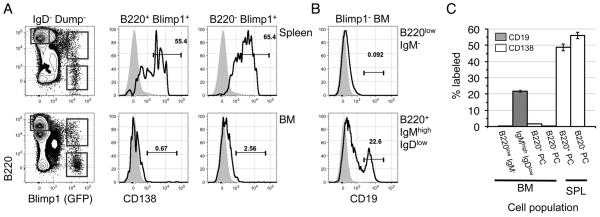
Figure 3. B220+ marrow plasma cells are not located in bone marrow sinusoids.
(A, B) B6.Blimp1+/GFP adults were given a single i.v. inoculation of 0.4 μg of PE-CD138 or PE-CD19. Two minutes later mice were sacrificed and BM and spleen cells stained with the indicated antibodies (excluding CD138 and CD19) before flow cytometric analysis of 2.5 × 10⁶ events. (A) Representative CD138 labeling of spleen (top) or BM (bottom) cells is shown. Left-most plots are pre-gated on viable Dump− IgD− cells as in Figure 1. Overlay histograms illustrate CD138 signal on the indicated PC subsets (black line), with the indicated B220high Blimp1/GFP− gates used to establish background signal (gray filled). (B) CD19 labeling for BM pro/pre-B cells (GFP− B220low IgM−) and immature (GFP− B220+ IgMhigh IgDlow) B cells. Gray filled curves are from mice that were not inoculated with PE-labeled antibodies. (C) Mean and SEM of % labeled cells within the indicated populations, with 3–5 mice per group summarized from two separate experiments.

