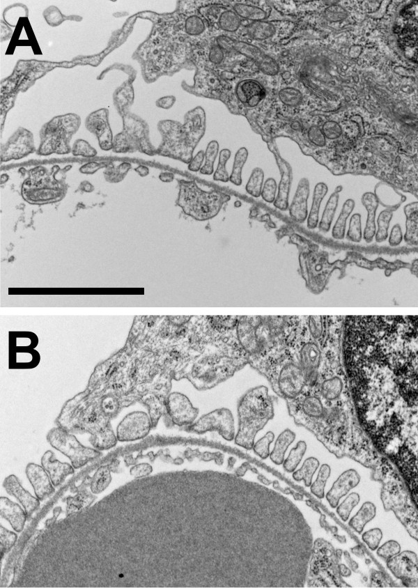
Figure 7.

Electron micrographs of glomeruli from WT and Clic4 null mice. A. Wild type. B. Clic4 null. In each, a section of a glomerular capillary is shown with the blood space below and the urinary space above, separated by basement membrane. The urinary surface of the basement membrane is lined with well-formed podocyte foot processes. The blood surface is lined with fenestrated endothelium. Scale bar represents 2 microns.
