Figure 7.

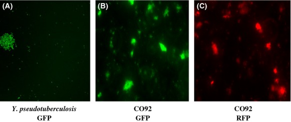
Fluorescence micrographs of (A) Yersina pseudotuberculosis GFP, (B) Y. pestis CO92 GFP, and (C) Y. pestis CO92 RFP strains with the fusions inserted into the chromosomal Tn7 site.

Fluorescence micrographs of (A) Yersina pseudotuberculosis GFP, (B) Y. pestis CO92 GFP, and (C) Y. pestis CO92 RFP strains with the fusions inserted into the chromosomal Tn7 site.