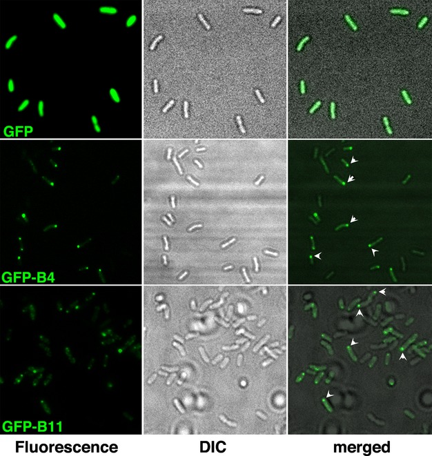
Figure 1.

Subcellular localization of GFP-VirB4 and GFP-VirB11. Subcellular location of GFP and its fusions with VirB4 or VirB11 was determined by epifluorescence microscopy. Left panels, fluorescence microscopy; middle, light microscopy; right, both together. GFP, GFP alone; GFP-B4, GFP-VirB4; GFP-B11, GFP-VirB11. A subset of polar foci is identified with arrowheads.
