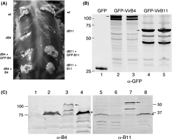
Figure 2.

DNA transfer activity and accumulation of GFP-VirB4 and GFP-VirB11 fusion proteins. (A) The ability of the fusion proteins to support tumor formation on Kalanchoe leaf was determined. Leaves were infected with the strains indicated. wt, wild-type Agrobacterium A348; dB4, A348ΔB4; dB11, A348ΔB11; GFP-B4, pADI15; GFP-B11, pADI16; B4, pvirB4; B11, pVirB11. (B,C) Accumulation of GFP-VirB4 and GFP-VirB11 was monitored by western blot assay following SDS-PAGE. Blots were probed with anti-GFP, anti-VirB4 or anti-VirB11 antibodies. An arrow identifies the full-length fusion protein. Numbers on left/right indicate the molecular mass (kDa) of marker proteins. The VirB4/VirB11-specific band in Figure2C is marked with an asterisk. (B) Lane 1, GFP; lanes 2 and 3, GFP-VirB4; lanes 4 and 5, GFP-VirB11. (C) Lanes 1 and 6, uninduced A348; lanes 2 and 5, induced A348; lane 3, GFP-VirB4; lanes 4 and 8, A348ΔB/pvirB; lane 7, GFP-VirB11.
