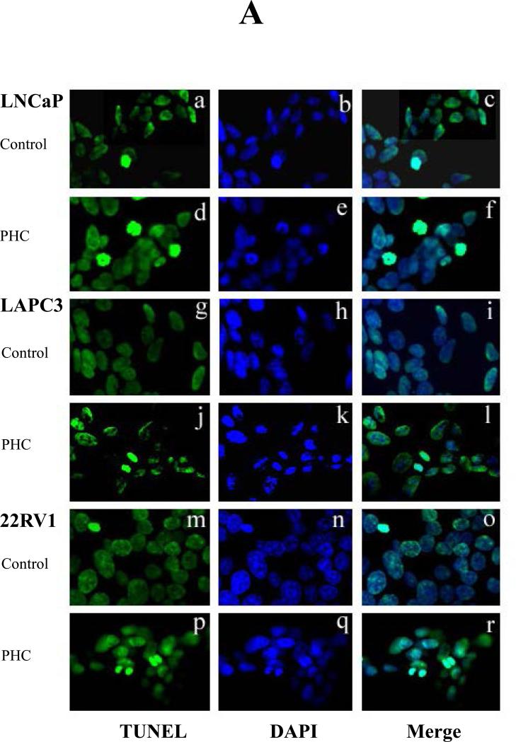
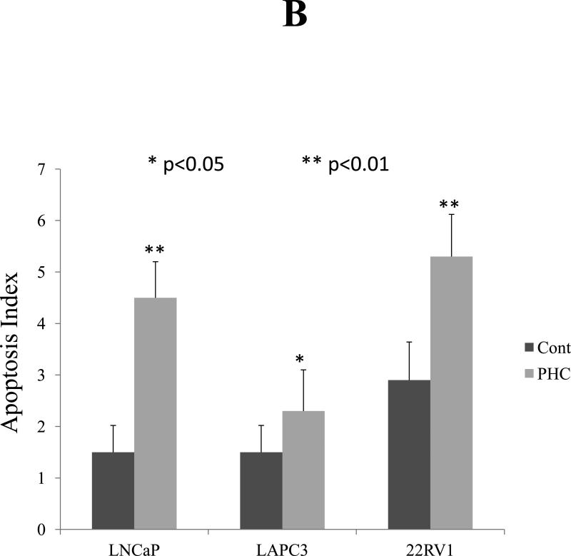
Figure 3. PHC Induces Apoptosis in Prostate Cancer Cells.
Three cells lines, LNCaP, LAPC3 and 22RV1, of prostate cancer were cultured on cover slips and treated with PHC (10 μg/ml) for 4 days. The PHC-induced apoptosis was examined by identifying DNA fragmentation with fluorescent TUNEL assay and counter-staining with DAPI. All apoptotic (TUNEL-positive) cell nuclei had bright green fluorescence under a fluorescence microscope. The result was expressed as an apoptotic index defined as the mean of TUNEL-positive cells counted per 400x field. P<0.05 indicated significant difference. Representative photomicrographs demonstrated the DNA fragmentation and dysmorphic nuclear condensation (Figure 3A), which were significantly increased in response to PHC treatment in all three cell lines (Figure 3A, d-f, j-l, p-r, Figure 3B).


