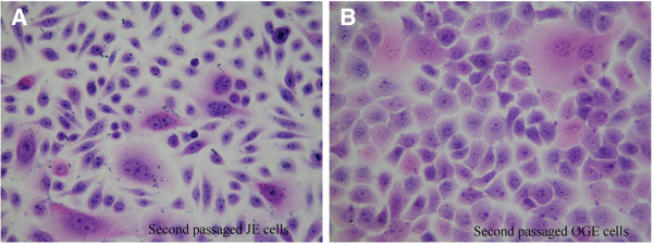
Figure 5.

H&E staining of JE and OGE cells (second passaged cells, H&E × 400). A. JE cells present non-uniform in size and morphology, scattered arrangement, large and deeply-stained nuclei and multiple nuclear divisions; B. OGE cells were uniform in size and shape, tightly arranged, and ‘paving stone-like’ keratinizing.
