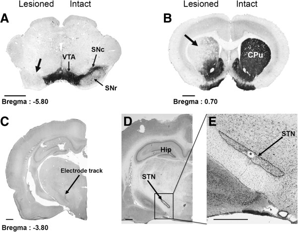
Figure 1.

Photographs of TH-immunostained coronal rat-brain sections at the nigral (A) and striatal (B) levels and of cresyl violet-stained coronal rat-brain sections at subthalamic (C, D and E) levels in 6-OHDA-lesioned rats. Note, on the lesioned side (left), the loss of dopaminergic cells in the SNc (A) and the loss of dopaminergic terminals in the striatum (B). Note also the correct implantation of the stimulation electrode within the STN (C, D, E). C, The arrow indicates the electrode track. E, The asterisk indicates the point of stimulation. CPu: Caudate Putamen; Hip, Hippocampus; SNc, Substantia nigra pars compacta; SNr, Substantia nigra pars reticulata; STN, Subthalamic nucleus; VTA, Ventral Tegmental Area. Scale bar, 0.75 mm.
