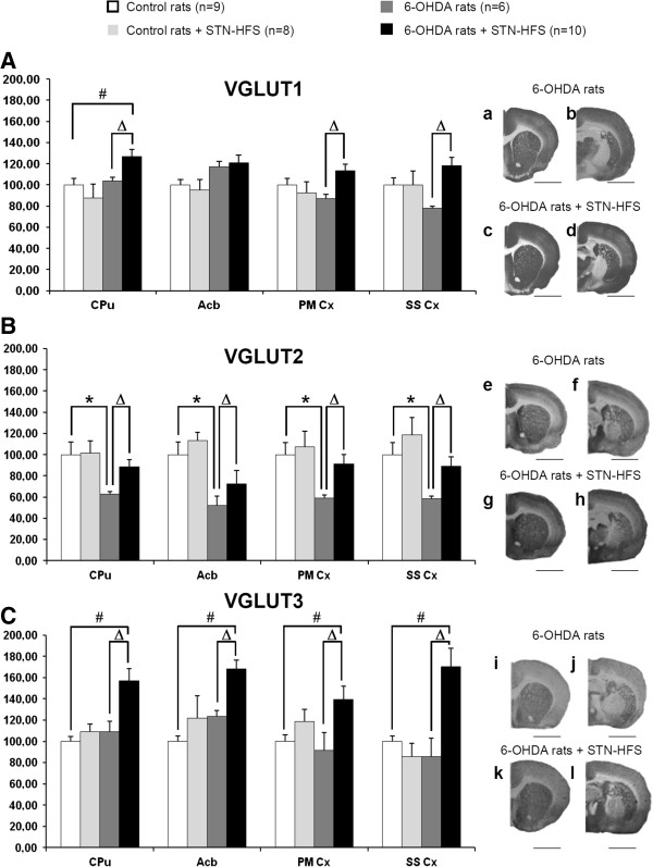
Figure 3.

Effects of 6-OHDA-lesion and STN-HFS on striatal and cortical VGLUT1-3 expression. A-C, Histograms show the mean ± standard error of the mean (SEM) of optical density values expressed as a percentage of values of control rats (non lesioned and non stimulated). Data were analyzed for each brain structure by Kruskal-Wallis tests with SigmaStat 3.1 software. Post-hoc analyses were carried out with the Dunn’s method. Kruskal-Wallis tests (VGLUT1, CPu, p = 0.024; Acb, p = 0.05; PMCx, p = 0.049; SSCx, p = 0.02), (VGLUT2, CPu, p = 0.009; Acb, p = 0.002; PMCx, p = 0.012; SSCx, p = 0.002), (VGLUT3, CPu, p = 0.003; Acb, p<0.001; PMCx, p = 0.043; SSCx, p = 0.002). a-l, Photographs of immunoradioautograms obtained by incubating coronal rat-brain sections with affinity-purified anti-VGLUT1 (a-d), anti-VGLUT2 (e-h) and anti-VGLUT3 (i-l) antisera and then with 125I-labeled anti-rabbit IgG. Acb, Accumbens nucleus; CPu, Caudate putamen; PM Cx, Premotor cortex; SS Cx, Somatosensory Cortex. *, controls vs 6-OHDA rats; #, controls vs 6-OHDA + STN-HFS rats; Δ, 6-OHDA rats vs 6-OHDA + STN-HFS rats: p < 0.05. Scale bar, 0.4 mm.
