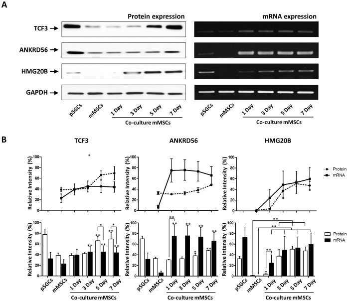
Figure 6. Quantitative analyses of ANKRD56, HMG20B and TCF3 expression using western blotting and RT-PCR.
A) Total protein lysate and mRNA samples isolated from the pSGCs derived from the submandibular gland tissue of 4 week-old B6 mice were used as a positive control. GAPDH protein was used for a loading control. Tcf3, hmg20B and Ankrd56 proteins were analyzed in pSGCs and co-cultured mMSCs. B) Densitometer analyses of salivary acinar cell markers, such as α-AMY, M3R and AQP-5, were analyzed in three independent replicates (p<0.05, one-way ANOVA with Bonferroni post-hoc test).

