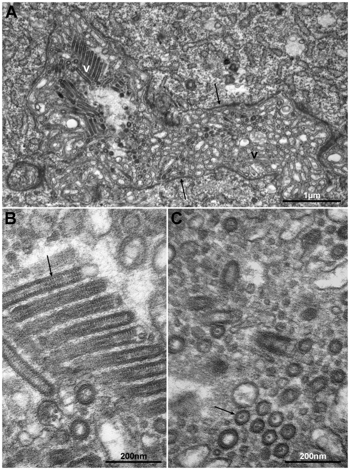
Figure 4. Section through a channel (lu) in a gland cell containing large amounts of virus-like particles (V).
Note the accumulation of electron dense material (arrows) on the inside of the cell membrane (A). The virus particles (arrow) are enveloped, rod-shaped or bacilliform, and appear as being cross-striated in tangential longitudinal sections (B). Transverse section through virus particles showing surrounding unit membrane and an electron dense core (C).

