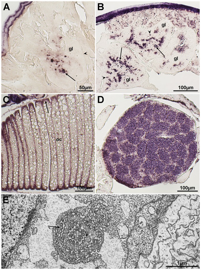
Figure 8. In situ hybridization for localization of LSRV genomes and mRNAs encoding the putative N protein.
In situ hybridization with an antisense probe targeting mRNA encoding the N protein of LSRV-No9 results in patches of staining (arrow) within an exocrine gland (gl), where the arrowhead is pointed at the gland capsule. These patches may represent viroplasm (A). A sense probe targeted at the LSRV-No127 genome induces coloring in or around gland (gl) secretory ducts, which are indicated by arrows. This may reflect viral budding through the cytoplasmic membrane and the presence of mature virions within the lumen of the duct, as shown by TEM (Figure 3 and 4). Patches of staining (arrowheads) in the cytoplasm may reflect viroplasm (B). Utilization of an antisense probe aimed at LSRV-No127 mRNA encoding the N protein, results in staining (arrow) of cytoplasm at the periphery of oocytes (oc), and within the ovary (figures C and D). TEM picture of putative virions budding (arrow) into the lumen of ER (E). It is not known if these spherical virus-like particles (arrow head) are connected to any of the two rhabdoviruses. Nucleus of the ovary cell (nu).

