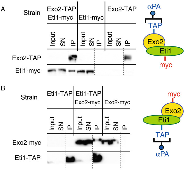
Figure 3.

The Exo2 and Eti1 proteins interact with each other. (A) Cells expressing Exo2-TAP, Eti1-myc, or both, were used for immunoprecipitation experiments with antibodies against protein A. Samples were analysed by Western blotting and probed with peroxidase anti-peroxidase complexes to detect the TAP tag (top panel), or with antibodies against the myc epitope (bottom panel). Equivalent amounts of proteins were loaded for the cell extract (Input) and the supernatant after purification (SN), and the immunoprecipitate (IP) was concentrated at least 5-fold with respect to the original extracts. All samples were run on the same gel. The dotted line indicates where a lane was removed from the image. Eti1-myc is only detected in the IP when Exo2-TAP is coexpressed. (B) As in A, but cells expressed Exo2-myc, Eti1-Tap, or both. Exo2-myc is specifically associated with Eti1-TAP. Note that myc-tagged proteins are detected more efficiently than TAP-tagged versions (probably due to the presence of 13 copies of the myc epitope). This explains why Exo2-myc, but not Exo2-TAP, can be detected in cell extracts (cf. panels A and B).
