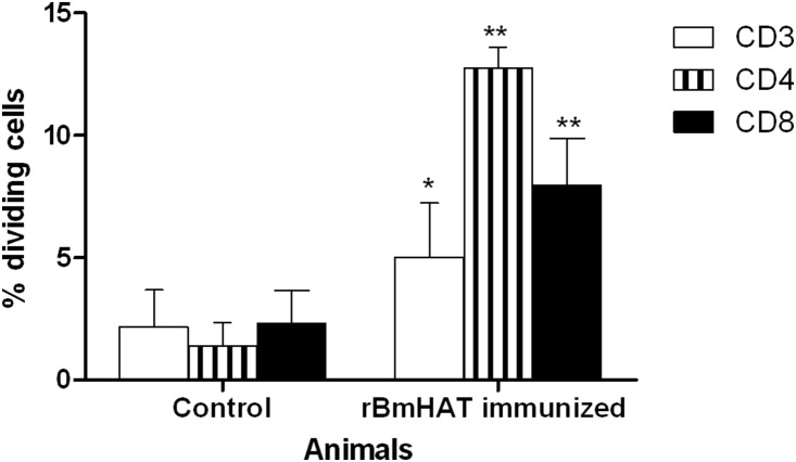
Figure 3. T cells proliferative responses in rBmHAT vaccinated macaques.
PBMC from immunized macaques isolated at 4 weeks post final immunization were stimulated with rBmHAT or medium only (negative control). Flow cytometry analysis for the loss of CFSE labeling (indicating cell proliferation) was performed for cells within the live T cell (CD3+) or CD4+ or CD8+ T cell subsets. Shown are the mean ± S.I and results (% proliferating cells) of the five macaques in each group. The frequency for each sample is the value obtained following subtraction of the medium control. Significant **P<0.001 and *P<0.05 proliferation of cells observed compared to controls.

