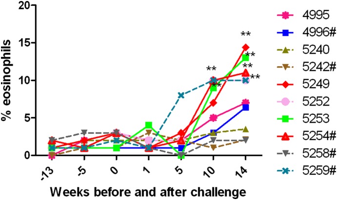
Figure 5. Percent of eosinophils in the peripheral blood of control and vaccinated macaques.
Percent of eosinophils were evaluated in the peripheral blood of macaque pre- and post- challenge. Following challenge there was an increase in the frequency of eosinophils in animals that were microfilaremic as determined by Knott’s technique. Each line represents the eosinophil count of individual macaque on weeks −13, −9, −5, −1 before challenge, on day of challenge (week 0) and at weeks 1, 5, 10, and 14 post challenges. The levels of eosinophils in Mf positive animals are represented by a solid line. Levels of eosinophils in Mf negative animals are represented as a dotted line. # Macaques immunized with rBmHAT. Significant at **P<0.001 high number of eosinophils.

