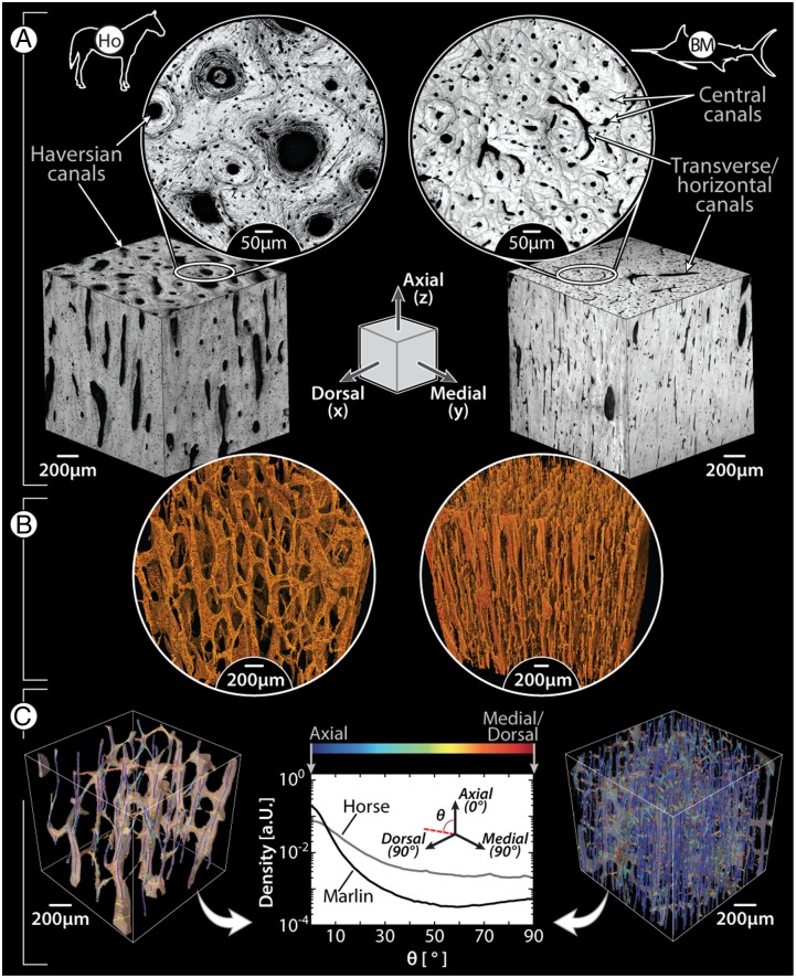
Fig. 4.
Comparison of vascular canal morphology in mammals and billfishes, using horse limb bone (Left) and blue marlin rostral bone (Right) as examples; the illustrated schematic in the center of A shows the anatomical orientation of all images, with the axial/longitudinal axis of the bones running from the top to bottom of the page. (A) Light microscopy images of the orthogonal faces of bone cubes (Inset images show higher magnification of the transverse plane, perpendicular to the axial direction); note the size difference between horse and blue marlin osteons and the presence of osteocytes in the horse image (small black dots surrounding each canal). (B) Volume-rendering of a microCT scan of each species’ canal network; note the comparatively large horizontal canals in blue marlin. (C) Volume-rendered skeletonization of canal networks, with canal segments color-coded according to orientation relative to the axial direction (blue, axial; red, perpendicular to axial, i.e., medial/dorsal). The frequency plot of these orientations illustrates that the majority of canals are oriented axially (0°) in both species; however, blue marlin also show a tendency toward a second peak at 90°, whereas horse exhibit more canal segments with intermediate angles (0° > θ > 90°). Horizontal canals in blue marlin (e.g., visible in A) are obscured by the extremely dense network of vertical canals in the volume renderings in B and C.

