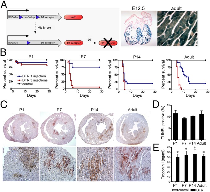
Fig. 1.
Age-dependent susceptibility to DT-induced cardiac injury. (A) Schematic of the DT cardiomyocyte injury model (Left). β-Galactosidase staining of E12.5 (Middle) and adult (Right) Rosa26R-LacZMlc2v-cre hearts. (B) Kaplan-Meier graphs of P1, P7, P14, and adult animals subjected to DT-induced cardiac injury. Black, control; blue, one DT injection; red, three DT injections. (C) TUNEL staining of P1, P7, P14, and adult animals after three DT injections. (D) Quantification of the percentage TUNEL-positive area in injured P1, P7, P14, and adult animals. (E) Troponin I serum levels immediately after three DT injections. *P < 0.05 compared with control.

