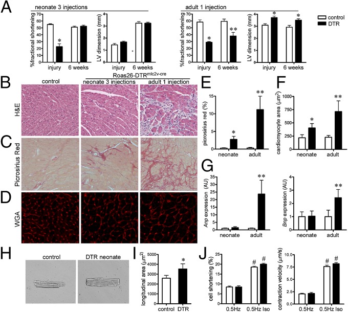
Fig. 4.
The neonatal heart functionally recovers after DT-induced cardiac injury. (A) Echocardiographic analysis of neonatal (three DT injections) and adult (one DT injection) hearts immediately after and 6 wk after injury. (B) H&E staining of surviving control and Rosa26-DTRmlc2v-cre hearts 6 wk after DT-induced cardiac injury. (C) Picrosirius red staining (fibrosis) 6 wk after neonatal and adult cardiac injury. (D) Wheat germ agglutinin (WGA) staining showing a cardiomyocyte cross-sectional area after cardiac injury. (E and F) Quantification of Picrosirius red staining (E) and cardiomyocyte cross-sectional area (F). (G) Anp and Bnp mRNA expression 6 wk after cardiac injury. (H and I) Longitudinal area of cardiomyocytes isolated from control and surviving Rosa26-DTRmlc2v-cre mice 6 wk after neonatal cardiac injury. (J) Cardiomyocyte cell shortening and contraction velocity at baseline and after isoproterenol treatment. (B and C) 20× objective. (D) 40× objective. *P < 0.05 compared with control; **P < 0.05 compared with all other groups; #P < 0.05 compared with cardiomyocytes not treated with isoproterenol.

