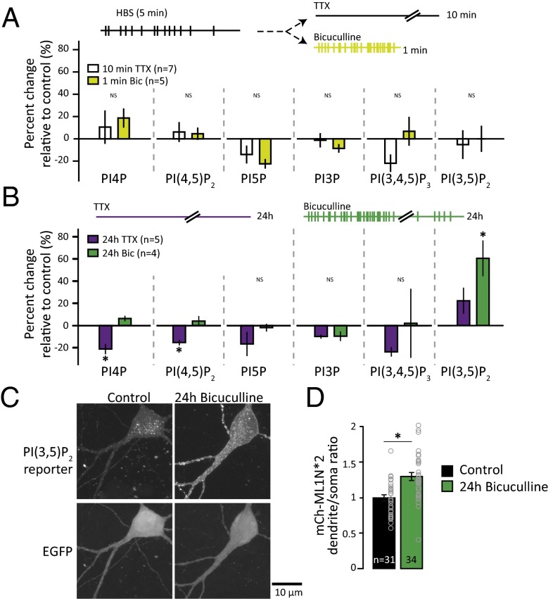
Fig. 3.
PI(3,5)P2 synthesis accompanies homeostatic synaptic weakening following prolonged hyperactivity. (A) Schematic of experimental design. Neurons were metabolically labeled with [3H]-inositol for 24 h. The media were replaced with Hepes-buffered saline (HBS). After 5 min, HBS was replaced with HBS + 2 μM TTX for 10 min or 50 μM bicuculline for 1 min and lipids were extracted. For each experiment, unstimulated controls (5 min of HBS) were collected and the mean (±SEM) PIP level after incubation with TTX or bicuculline is expressed as a percentage of the control (5 min of HBS). (B) Schematic of experimental design. Neurons were metabolically labeled with [3H]-inositol for 24 h in the presence of DMSO control, 2 μM TTX, or 50 μM bicuculline. The mean (±SEM) percent change in PIP levels relative to control from neurons incubated with 2 μM TTX (n = 5) or 50 μM bicuculline (n = 4) for 24 h is shown. The level of PI4P decreased after 24 h of TTX [percent change: −21.26 ± 1.77%; one-way ANOVA: F(2,11) = 15.2, P = 0.0007]. The level of PI(4,5)P2 decreased after 24 h of TTX [percent change: −15.34 ± 2.45%; one-way ANOVA: F(2,11) = 6.54, P = 0.014]. The level of PI(3,5)P2 increased after 24 h of bicuculline [percent change: 60.49 ± 16.05%; one-way ANOVA, F(2,11) = 7.13, P = 0.01]. (C) Representative images of neurons expressing the PI(3,5)P2 reporter, mCherry-ML1N*2, and GFP. (D) Mean (±SEM) ratio of dendritic mCherry-ML1N*2 fluorescence to soma mCherry-ML1N*2 fluorescence. The dendritic-to-soma ratio of mCherry-ML1N*2 intensity increased after 24 h of bicuculline [control: 1.0 ± 0.04, 24 h of bicuculline: 1.30 ± 0.06; t test: t(63) = 4.12, P = 0.0001]. *P < 0.05.

