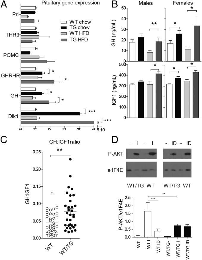
Fig. 5.
Dlk1-overexpressing mice have elevated pituitary growth hormone secretion as a result of local resistance to IGF1 feedback. (A) RT-qPCR of anterior pituitary genes in WT and WT/TG animals fed a chow or HFD at 24 wk (fasted for 16 h, 70C mice), relative to WT chow = 1.0. WT and WT/TG, n = 7 per genotype. Prl, prolactin. (B) Serum GH (Upper) and IGF1 (Lower) in 16-h fasted chow and HFD-fed mice, measured between 1000 hours and 1200 hours, n = 6–15 per sex per genotype. *P < 0.05, **P < 0.01 by Student's t-test. (C) Ratio of GH:IGF1 levels from all WT and WT/TG mice above (n = 33 WT and 32 WT/TG, males and females combined because no significant difference was found between them). **P < 0.01 by Mann–Whitney U test. (D) Western blot of AKT phosphorylation after IGF (I) or IGF1 plus DLK1 (ID) stimulation of pituitary explants from WT and WT/TG mice (Upper). The level of phosphorylation was normalised to total protein levels and charted (Lower), compared by one-way ANOVA with Bonferroni’s multiple comparison test, **P < 0.01, ***P < 0.001.

