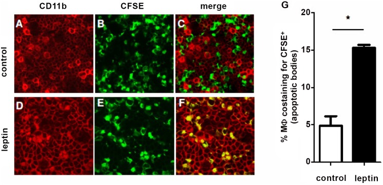
Figure 2. Leptin promotes phagocytosis by lupus macrophages in vivo.

NZB/W mice were injected i.p. with thioglycolate prior to injection of either vehicle (A–C) or 2 µg/g leptin (D–F) at 12-h intervals. After 72 h, 1×10⁶ CFSE-positive apoptotic cells were injected i.p. After 30 min, uptake of apoptotic cells by CD11b+ macrophages (PE) in peritoneal fluid of recipient animals was visualized ex vivo by confocal microscopy as colocalization of PE-positive macrophages (red) and CFSE-positive (green) apoptotic bodies within the same cell (yellow). Representative of three experiments (n = 6 per group). Original magnification: 10x. (G) Cumulative flow cytometry of peritoneal macrophages costaining for apoptotic cells. *p<0.01 vs control.
