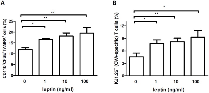
Figure 5. Increased macrophage uptake of apoptotic cells induced by leptin promotes proliferation of self antigen-reactive T cells.

TAMRA-labeled apoptotic bodies containing OVA (see Methods) were co-cultured with CFSE-labeled macrophages for 2 h, followed by staining with APC-labeled anti-CD11b Ab and flow cytometry. (A) Frequency of TAMRA+CFSE+ CD11b+ cells (macrophages positive for OVA-containing apoptotic bodies) in OVA-TCR transgenic mice (DO11.10) after culture in the presence of scalar doses of leptin. (B) Flow cytometry staining with clonotype-specific KJ1.26 Ab (OVA-specific TCR) 48 h after co-incubation of OVA-immunized DO11.10 (OVA-TCR transgenic) mouse splenocytes with macrophages containing OVA-loaded apoptotic bodies (TAMRA+). *p<0.05, **p<0.01 vs control (n = 5).
