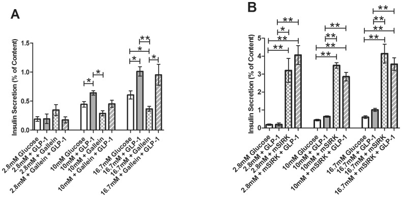
Figure 6. Percent of insulin content secreted from intact islets after static incubation at 2.8, 10, and 16.7 mM glucose with and without treatment.
Untreated control samples are shown in white. A, Percent of insulin content secreted at 2.8, 10, and 16.7 mM glucose concentrations in the presence and absence of GLP-1 (20 nM, light gray), the Gβγ inhibitor, gallein (10 µM, checked), or combination treatment with gallein and GLP-1 (striped). B, Percent of insulin content secreted at 2.8, 10, and 16.7 mM glucose concentrations with GLP-1 (20 nM, light gray), the Gβγ-activating peptide mSIRK (30 µM, checked), or combination treatment with mSIRK and GLP-1 (striped). Data are the mean ± S.E. n = 4–19. *(p<0.05) and **(p<0.001) indicate significance compared to untreated control, GLP-1 only, or gallein alone.

