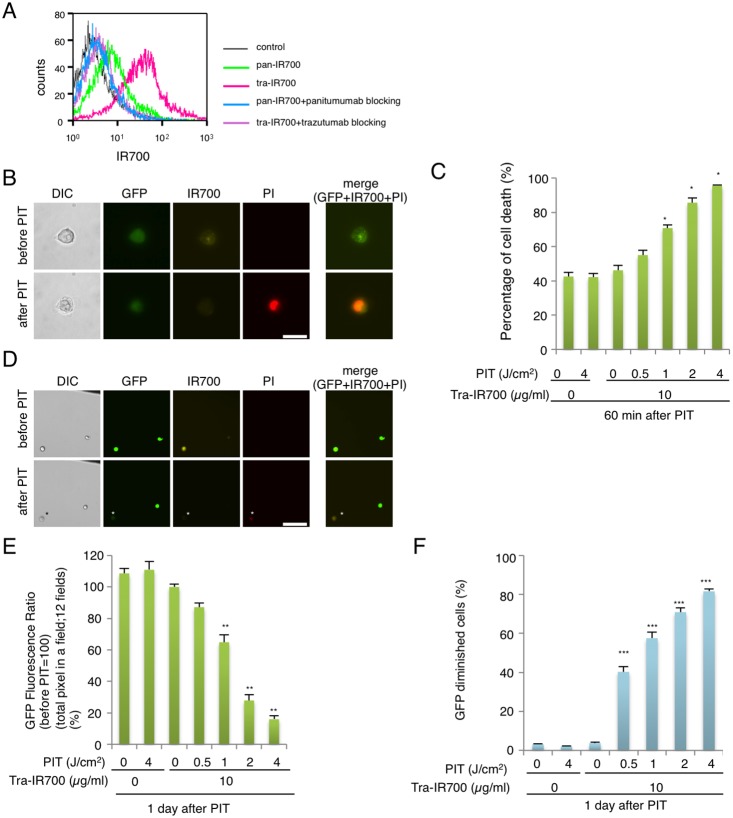
Figure 1. Confirmation of HER2 expression as a target for PIT in N87-GFP cells, and evaluation of in vitro PIT.
(A) Expression of HER1 and HER2 in N87-GFP cells was examined with FACS. HER2 was overexpressed more than HER1. Specific binding was demonstrated by antibody blocking. (B) N87-GFP cells were incubated with tra-IR700 for 6 hr and observed by microscopy (Before and after irradiation of NIR light; 2 J/cm2). Necrotic cell death was observed upon excitation with NIR light (after 30 min). Bar = 25 µm. PI staining showed the membrane damage. (n = 4, *p<0.001, vs. untreated control, Student’s t test) (C) Membrane damage induced by PIT was measured with the dead cell count using PI staining, which increased in a manner dependent on the light dose. (D) N87-GFP cells were incubated with tra-IR700 for 6 hr and irradiated with NIR-light (0.5 J/cm2). GFP-fluorescence intensity decreased in dead cells but was unchanged in living cells at 1 day after PIT (*). Bar = 200 µm. The black line at right upper corner was the marker to determine the position of observation. (E) Diminishing GFP-fluorescence intensity at 1 day after PIT occurred in a manner dependent on the light dose (total pixel of GFP fluorescence in the same) (n = 12 fields) (**P<0.0001, vs. untreated control, Student’s t test). (F) Diminishing GFP fluorescence intensity induced by PIT, as measured by FACS, confirms a NIR-light dose-dependence. (n = 4, ***P<0.0001, vs. untreated control, Student’s t test).

