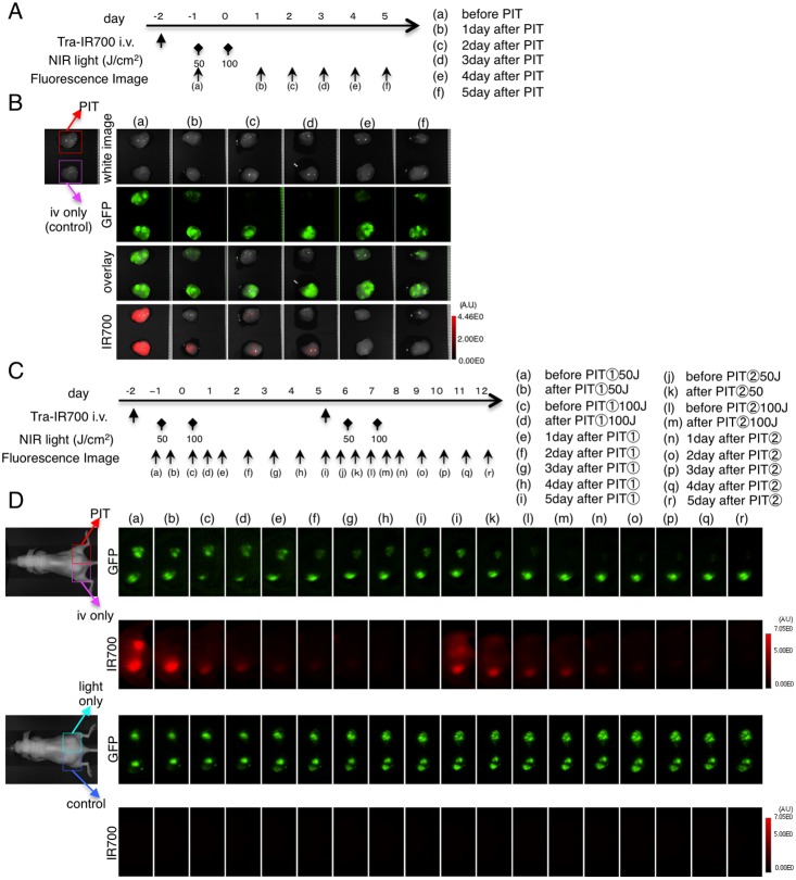
Figure 4. Ex vivo GFP fluorescence imaging after repeated PIT in bilateral N87-GFP flank model.
(A) PIT regimen. Fluorescence images of ex vivo tumors were obtained at each time point as indicated. (B) Ex vivo fluorescence imaging of N87-GFP tumor in response to PIT. Fluorescence changes are seen with both GFP and IR700 fluorescence in response to PIT. (C) Fluorescence images were obtained at each time point as indicated. (D) In vivo fluorescence real-time imaging of bilateral flank tumor bearing mice in response to repeated PIT. GFP-fluorescence intensity of the tumor was almost eliminated after the second PIT.

