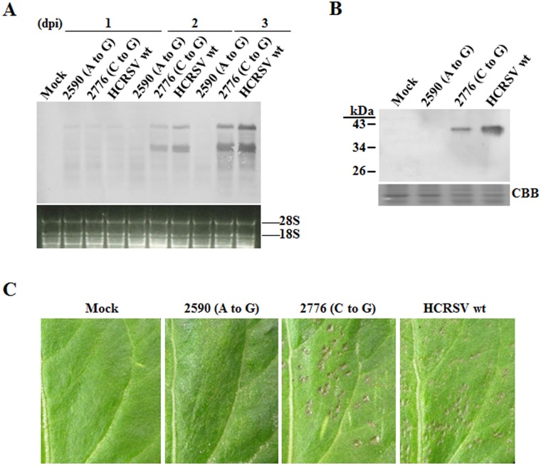
Figure 3. HCRSV viral RNA and CP accumulation in inoculated kenaf cotyledons.
In vitro transcripts (0.5 µg for each cotyledon) were inoculated onto kenaf cotyledons and the inoculated cotyledons were collected at different time points. (A) Northern blot analysis of viral RNA. Total RNA (5 µg each) extracted from cotyledons at 1, 2 and 3 days post inoculation (dpi), respectively, was used for viral RNA detection. (B) Western blot analysis of viral CP. Total protein extracted from cotyledons at 4 dpi was used for HCRSV CP detection by western blot. (C) Observation for local lesions in inoculated kenaf cotyledons at 4 dpi.

