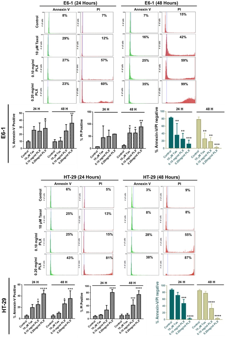
Figure 3. Quantification of Cell Death Induction Following PLX Treatment.
Image-Based cytometry was used to quantify apoptotic induction (% Annexin V positive), followed by necrosis (% PI positive) in E6-1 and HT-29 cells following PLX treatment. The lack of annexin V or PI staining was used as an indication of live cells following treatment (%Annexin V/PI negative cells) (*P<0.05, ** P<0.003, ***P<0.0001). (E) To further confirm the induction of apoptosis.

