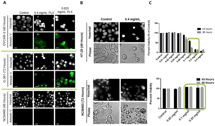
Figure 5. PLX Selectively Targets Cancer Cells for Apoptosis Induction.
Subsequent to treatment with PLX, cells (Ovarian; OVCAR-3, Melanoma; G-361 and Normal Colon Epithelia cells (NCM460) were stained with Hoechst to characterize nuclear morphology and Annexin-V to detect apoptotic cells (A) and cellular morphology by phase contrast microscopy (B); Images were taken at 400× magnification on a fluorescent microscope. Scale bar = 15 µm. (C) Following PLX treatment, HT-29 colorectal cancer cells and non-cancerous NCM460 cells were incubated with WST-1 cell viability dye for 4 hours and absorbance was read at 450 nm and expressed as a percent of the control. Values are expressed as mean ± SD of 3 independent experiments. **P<0.0001.

