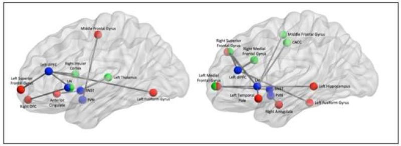
Fig 6.
Summarized findings showing differences in functional connectivity between non-anxious participants and elderly GAD during worry induction (left) and during worry reappraisal (right)
Legend: In blue – the four seeds (LAI=left anterior insula, dlPFC=dorso-lateral prefrontal cortex, BNST=bed nucleus of stria terminalis, PVN=paraventricular nucleus. In red-regions of interest that had greater connectivity with the seed for GAD than for non-anxious participants. In green: regions of interest that had greater connectivity with the seed for non-anxious participants than for GAD.

