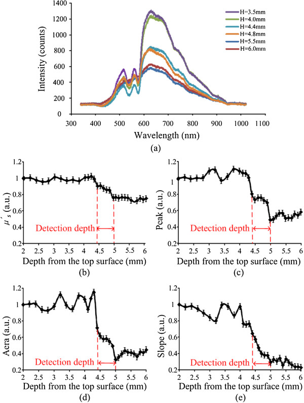
Figure 7.

Experiment results. (a) 6 NIRs spectrometer: H represents the depth from the top surface (b) the change of the μ ′ s (c) the change of the area (d) the change of the peak (e) the change of the slope. In Figure 5(a), the dark blue and red lines represent the measurement points in compact bone, the green and purple lines represent the measurement points in spongy bone, and the blue and orange lines were just between them, which mean the points in alarm region.
