
Figure 8.
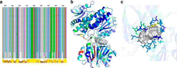
Sequence and structural level details of Rv3436c (glmS). a) Snapshot of MSA of this gene over the Mycobacterium genus is shown. Conservation scores for each residue in a protein are calculated using in-house algorithm; b) shows the protein structure of Rv3436c on which the predicted conserved residues are mapped. A colour scale (from blue to red denoting highest to the least) is used. c) One of the binding pockets of Rv3436c is shown and conserved residues that lie in the site are indicated. The pocket is shown in CPK representation and the residues forming the pockets are shown as stick model.