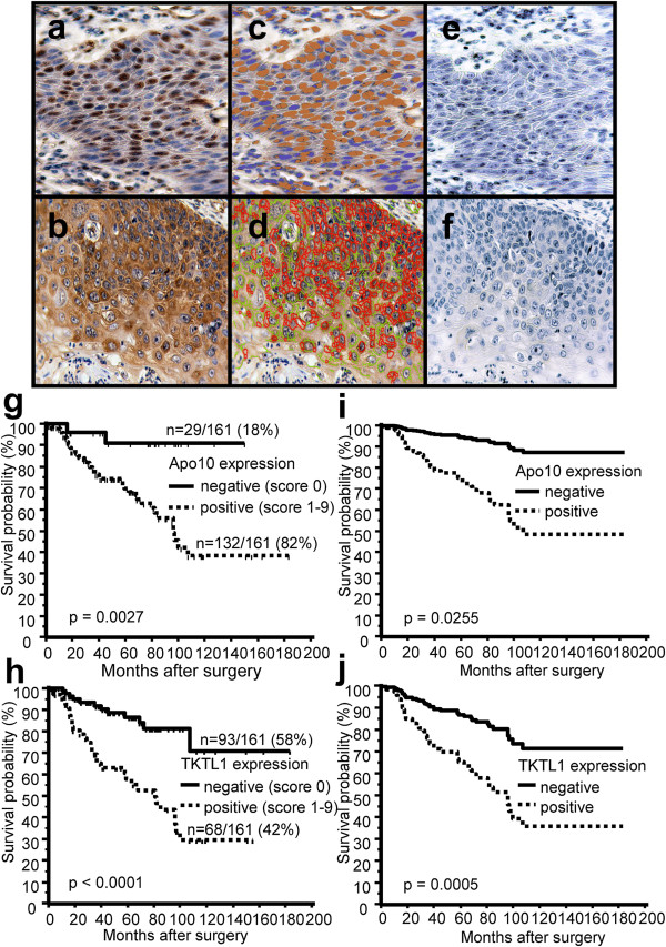
Figure 1.

Immunohistochemical single staining of Apo10, TKTL1 and survival curves of OSCC patients measured by Apo10 and TKTL1 expression. Brown chromogen color (3,3′-Diaminobenzidine, DAB) indicates positive Apo10 staining (a, nuclear staining pattern) and positive TKTL1 expression (b, cytoplasmic staining pattern), the blue color shows the nuclear counterstaining by hematoxylin. Pseudo-colored images (c, d) show the staining components of computer-assisted quantitative analysis in Apo10+ and TKTL1+ tumor cells. Computer-assisted light brown label (c) indicates positive Apo10 staining and the computer-assisted light blue label marks the nuclei counterstained with hematoxylin. Computer-assisted red label (d) indicates strong or complete TKTL1 staining, the green label (d) indicates weak or incomplete staining. Apo10 staining is abolished after incubation with immunogenic peptide (e). Representative image of IgG control (f) shows no staining. Original magnification: ×200-fold. Kaplan-Meier (g, h, left panel) and Cox-regression (i, j, right panel) survival curves for disease-free survival (DFS) stratified by positive Apo10 and TKTL1 expression (Apo10+, TKTL1+, dashed lines) and negative Apo10, TKTL1 expression (Apo10-, TKTL1-, solid lines). In univariate Kaplan-Meier analysis positive Apo10 (g) and TKTL1 (h) expression is significantly associated with poorer survival. The times of the censored data are indicated by short vertical lines. Multivariate Cox-regression analysis shows positive Apo10 (i) and TKTL1 (j) expression as significant independent adverse prognostic factors.
