
Figure 1.
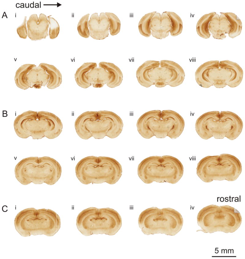
Expression of mGluR2 in the cortex of the developing mouse brain. Slides were scanned on a Nanozoomer-XR slide scanner. Each panel (A-C) depicts images scanned from one slide, with the sections mounted caudorostrally from left to right (Ai-viii, Bi-viii, Ci-iv). (A) In caudal sections, intense labeling for mGluR2 is found in the entorhinal cortical areas located in the ventral regions, as well as limbic structures. Labeling of the sensory cortical areas is evident in the dorsal cortical regions. (B) The pattern of labeling in the sensory cortical areas continues in sections containing the thalamus. Strong labeling is also seen in the thalamic reticular nucleus and moderate labeling in the basolateral amygdala. (C) In the rostral most sections, sensory cortical areas are still strongly labeled, but ventral cortical areas are very weakly labeled, particularly the piriform cortex. Additional labeling is found in the caudate putamen, hippocampal commissure, and retrosplenial gyrus.