
Figure 3.
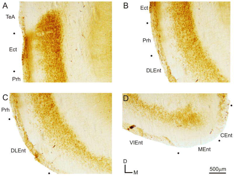
Expression of mGluR2 in caudoventral cortical areas. Higher magnification images of the ventral cortical areas from the section depicted in Figs. 1Aiii and 3A. (A) Labeling in the temporal association cortex (TeA), ectorhinal cortex (Ect), and perirhinal cortex (Prh). (B) Expression of mGluR2 in Ect, Prh, and dorsolateral entorhinal cortex (DLEnt). (C) Prh and DLEnt labeling of mGluR2. (D) Ventral intermediate entorhinal cortex (VIEnt), medial entorhinal cortex (MEnt), and caudomedial entorhinal cortex (CEnt) labeling pattern.