Figure 5.

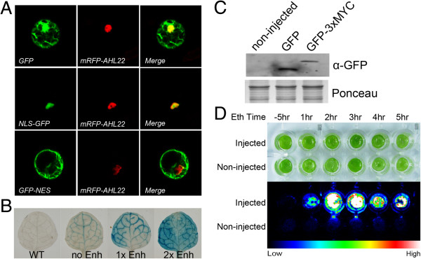
The verification of BioVector. A, Analysis of signal peptides. The expression constructs, Fu39-2-35S::NLS:GFP, Fu39-2-35S::GFP:NES, or Fu39-2-35S::GFP, respectively, were co-transfected with a nuclear protein marker (AHL22, [31]) construct (35S::mRFP-AHL22) into Arabidopsis protoplasts, and the fluorescence signal was observed under a confocol microscope after 14 hours incubation. B, Analysis of promoter activity. The constructs of Fu39-2-SUC2::GUS, Fu39-2-SUC2:Enh::GUS, Fu39-2-SUC2:2xEnh::GUS were transformed into Arabidopsis (Col). T1 transgenic plants for each construct were analyzed with GUS staining. C, Detection of tagged proteins. The Fu39-2X35S::GFP and Fu39-2x35S::GFP:3xMyc expression constructs were respectively introduced into Arabidopsis protoplasts, and the protein was extracted, subjected to SDS-PAGE, and then probed by anti-GFP antibody on a western blot. D, Ethanol induced expression of LUC. Fu39-8-Ub::LUC was infiltrated into 3-week-old N. benthamiana leaves mediated by Agrobacterium. The leaf disc was harvested on day 3 after infiltration and incubated in 1/2 MS liquid medium containing 2% (v/v) ethanol for indicating hours. Then the luciferin was added into the medium to a final concentration at 100 μM, and the bioluminescence signal was imaged by a CCD camera (Princeton). The bright field and luciferase imaging were respectively shown in the upper and lower panel. All experiments were carried out with at least three biological replicates.
